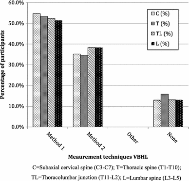
Method 1 anteriormiddle column vertebral body compression ratio vbcr avhpvh method 2 anterior vertebral body. Previously researchers used different measurement methods to assess the degree of vertebral body collapse by using posterior wall height as the reference vertebral body compression ratio vbcr or the percentage of anterior height compression pahc which uses the mean height of segments adjacent to a healthy vertebral body as the reference value 8 11.
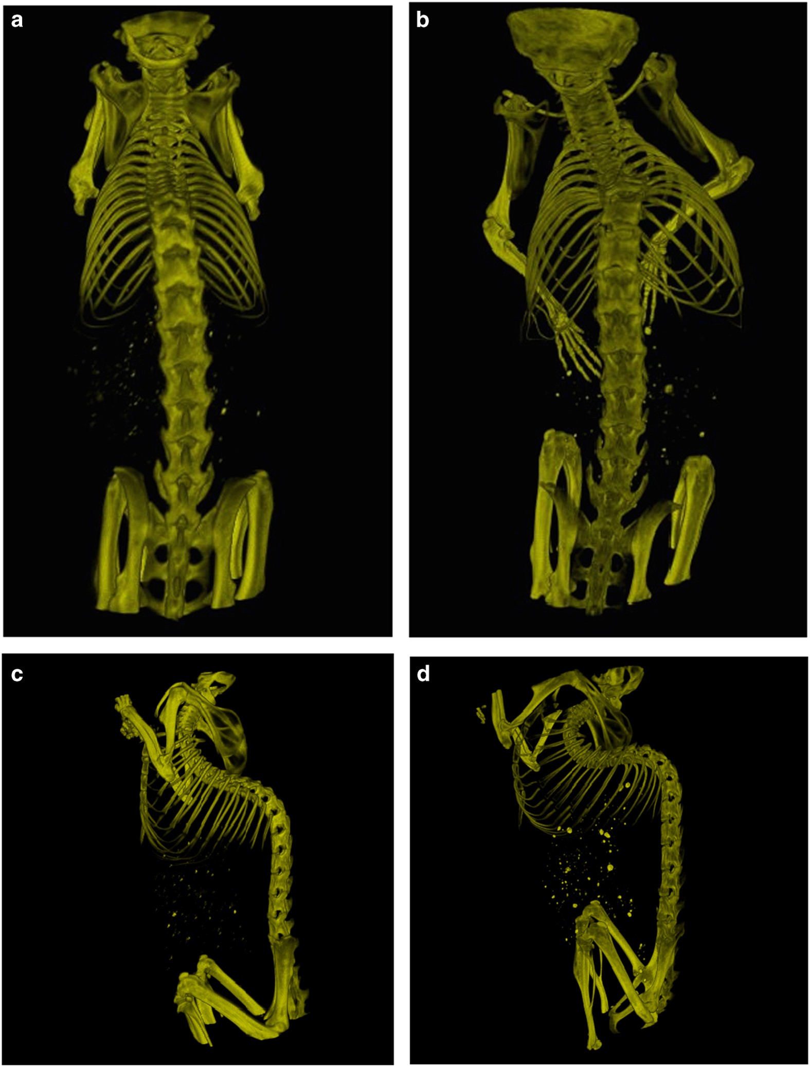
Development Of Spinal Deformities In The Tight Skin Mouse
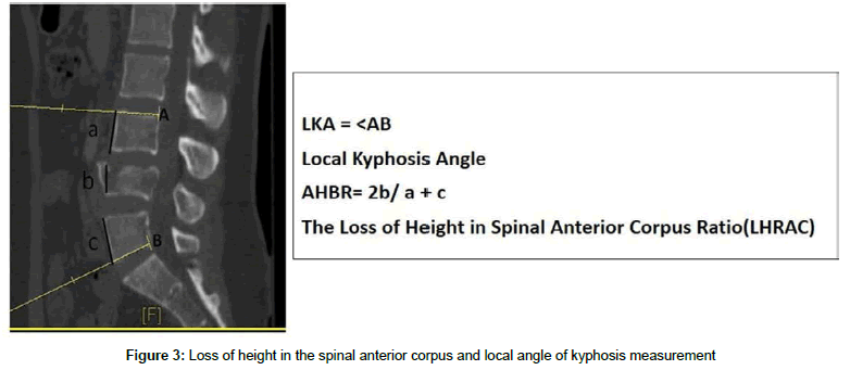
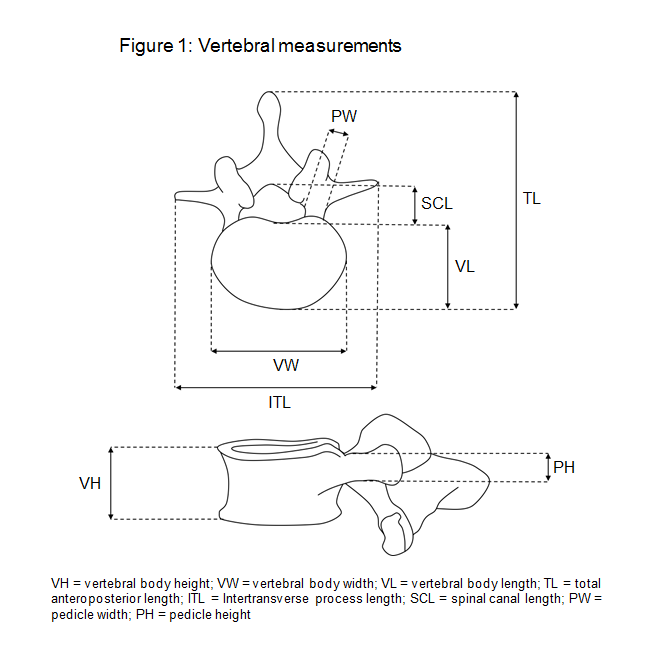
Vertebral body height measurement. The average dorsal t12v body height was 2125164 mm in the control group and 2011149 mm in the lvf group. Vertebral measurements measurements of vertebral body height were taken from the midline of each centra c3l5 at the point of maximum height. This study used previous measurement methods to assess the degree of vbhl and ka compare and examine differences between various measurement methods and examine the correlation between relevant measurement parameters and intravertebral cleft ivc in the vertebral body. For compression fracture vertebral body height loss vbhl and kyphotic angle ka are two important imaging parameters for determining the prognosis and appropriate treatment. The average ventral t12v body heights were 1951154 mm and 1762195 mm respectively. The lvf group had significantly lower dorsal and ventral t12v body heights both p0001.
Degenerative disk disease in the spine results in loss of disk height. Surveyed measurement techniques for assessing vertebral body height loss. An automated method for precise measurement of vertebral body height and intervertebral disk height using computed tomography vertebral fractures due to osteoporosis result in loss of vertebral height.
Gallery of Vertebral Body Height Measurement