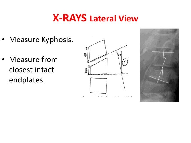
Along the length of the spine the vertebrae change to accommodate different needs related to stress and mobility. Measurement of kyphosis and vertebral body height loss in traumatic spine fractures.

Radiographic Measurement Of Vertebral Body Height In Sagittal
Measurement of vertebral body. Epub 2016 aug 6. It is a thick bony structure which provides strength to the spine and protection for the spinal cord. Method 1 anteriormiddle column vertebral body compression ratio vbcr avhpvh method 2 anterior vertebral body compression percentage avbc v2v1 v32 100 avh anterior. The size of the vertebral bodies increases down the spine as the size and weight of the body it has to support above it increase. In the human vertebral column the size of the vertebrae varies according to placement in the vertebral column spinal loading posture and pathology. The column runs from the cranium to the apex of the coccyx on the posterior aspect of the body.
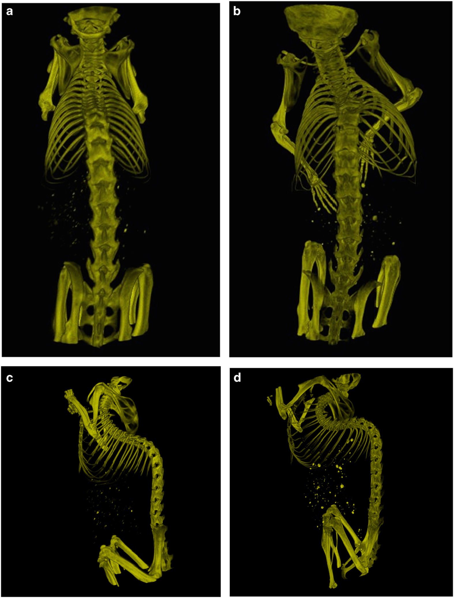
Five or in some cases six vertebrae make up the lumbar spine which provides support for much of the upper body and is rather flexible. Anterior middle and posterior vertebral body heights of all segments from t4 to l4 were measured interactively using mxa software and qm from the spinal radiographs and compared with direct measurements derived using digital callipers following cadaveric dissection. In the measurement of vertebral body heights the accuracy decreased at vertebral levels where the images of thoracic vertebral bodies are superimposed upon by the shadows of cardiovascular organs. A compression fracture usually heals in eight to ten weeks. The vertebrae are the bones which make up the spinal column in humans and other vertebratesthe human body has 33 vertebrae 24 of which make up the spinethe vertebral body is the largest part of each vertebra. The vertebral column also known as the backbone or the spine is a column of approximately 33 small bones called vertebrae.
Lumbar vertebrae are larger than the thoracic or cervical. The purpose of this study is to perform quantitative measurement based on the standardized uptake value suv of the uptake of tc 99m methylene diphosphonate mdp in the normal vertebrae using a single photon emission tomography spectcomputed tomography ct scanner. The vertebral body is the large anterior cylindrical portion that is predominantly responsible for bearing the weight of the spine and body above it. It contains and protects the spinal cord.
Gallery of Measurement Of Vertebral Body